


Interactive CT & MRI Anat.Lite

คำอธิบายของInteractive CT & MRI Anat.Lite
★Lite version★
This is the free Lite version of "Interactive CT and MRI Anatomy".
The function is restricted.
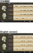
You can only see the transverse CT images of the head.
Please check the operation before purchasing the full version.
★ Details ★
This application is developed for medical students, interns, residents, doctors, nurses, and radiology technicians to understand the essential anatomical terms of the body.
You can learn anatomy by answering the terms by step-to-step questions using a total of 241 CT and MRI images.
A total of 17 images of 3D-CT, MRA and plain X-ray film(particularly the extremities) are included as references.
Other reference images include coronary artery segments defined by the American Heart Association(AHA), pulmonary segments, and liver segments(according to Couinaud classification).
You can enlarge all the images by simple manipulation.
★ Major functions ★
There are 4 major functions.
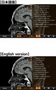
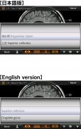
-1) Anatomical mode
Anatomical terms are overlaid on the images.
It can be used as the anatomical atlas.
-2) Quiz mode type 1
You select the part of the image by using anatomical term.
Questions will basically appear randomly.
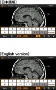
-3) Quiz mode type 2
You select the anatomical term by the part of the image.
Questions will basically appear randomly.
-4) Index
You can find the specific images by using anatomical terms.
★ Intended users ★
-Medical students
-Interns and residents
-Doctrors
-Nurses
-Radiology technicians
-All those who are intrested in CT and MRI anatomy
★ Images(a total of 258 images) ★
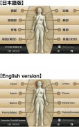
Images basically include horizontal, coronal, and sagital planes.
-Head(36 images including CTA and 3D-CT)
-Neck(24 images)
-Spine(19 images including plain X-ray films)
-Chest(61 images including 3D-CT images)
-Abdomen (37 images)
-Pelves: male (9 images)
-Pelvis: female (11 images)
-Extremities (shoulder, hand, elbow, hip joint, knee, foot) (61 images including plain X-ray films)
Editors
Toshiaki Nitori, M.D. (Professor of Radiology, Kyorin University, School of Medicine)
Yasuo Sasaki, M.D. (Manager of diagnostic radiology, Iwate Prefectural Central Hospital)
</div> <div jsname="WJz9Hc" style="display:none">★★รุ่น Lite
นี้เป็นรุ่น Lite ฟรีของ "อินเตอร์แอคที CT และ MRI กายวิภาคศาสตร์"
ฟังก์ชั่นจะถูก จำกัด
คุณจะเห็นเฉพาะภาพขวาง CT ของศีรษะ
กรุณาตรวจสอบการดำเนินการก่อนที่จะซื้อเวอร์ชันเต็ม
★★รายละเอียด
โปรแกรมนี้ถูกพัฒนาขึ้นสำหรับนักศึกษาแพทย์ฝึกหัดที่อาศัยอยู่, แพทย์, พยาบาล, และช่างเทคนิครังสีที่จะเข้าใจเงื่อนไขทางกายวิภาคที่สำคัญของร่างกาย
คุณสามารถเรียนรู้กายวิภาคโดยการตอบแง่คำถามโดยขั้นตอนในการใช้ขั้นตอนทั้งหมด 241 CT และ MRI ภาพ
รวม 17 ภาพ 3D-CT, MRA และธรรมดาฟิล์มเอ็กซ์เรย์ (โดยเฉพาะขา) ซึ่งรวมถึงการอ้างอิง
ภาพอ้างอิงอื่น ๆ ได้แก่ กลุ่มหลอดเลือดหัวใจที่กำหนดโดยสมาคมหัวใจอเมริกัน (AHA) ส่วนปอดและกลุ่มตับ (ตามการจัดหมวดหมู่ Couinaud)
คุณสามารถขยายภาพได้ทั้งหมดโดยการจัดการที่ง่าย
★★ฟังก์ชั่นที่สำคัญ
มี 4 ฟังก์ชั่นที่สำคัญคือ
-1) โหมดกายวิภาค
แง่กายวิภาคจะวางทับบนภาพ
มันสามารถใช้เป็นแผนที่ทางกายวิภาค
-2) ประเภทโหมดแบบทดสอบ 1
คุณเลือกส่วนของภาพโดยใช้ระยะทางกายวิภาค
คำถามที่พื้นจะปรากฏแบบสุ่ม
-3) ประเภทโหมดแบบทดสอบ 2
คุณสามารถเลือกระยะทางกายวิภาคโดยเป็นส่วนหนึ่งของภาพ
คำถามที่พื้นจะปรากฏแบบสุ่ม
-4) ดัชนี
คุณสามารถค้นหาภาพที่เฉพาะเจาะจงโดยใช้เงื่อนไขทางกายวิภาค
★★ผู้ใช้ตั้งใจ
นักเรียนทางการแพทย์
-Interns และผู้อยู่อาศัย
-Doctrors
-Nurses
ช่าง -Radiology
พักผู้ที่กำลัง intrested ใน CT และ MRI กายวิภาคศาสตร์
★ภาพ (รวม 258 ภาพ) ★
ภาพรวมโดยทั่วไปในแนวนอนเวียนและ sagital เครื่องบิน
-Head (36 ภาพรวมทั้ง CTA และ 3D-CT)
-Neck (24 ภาพ)
-Spine (19 ภาพรวมทั้งหนังเอ็กซ์เรย์ธรรมดา)
-Chest (61 ภาพรวมทั้งภาพ 3D-CT)
-Abdomen (37 ภาพ)
-Pelves: ชาย (9 ภาพ)
-Pelvis: หญิง (11 รูป)
-Extremities (ไหล่, มือ, ข้อศอก, ข้อต่อสะโพกหัวเข่าเท้า) (61 ภาพรวมทั้งหนังเอ็กซ์เรย์ธรรมดา)
บรรณาธิการ
Toshiaki Nitori, แมรี่แลนด์ (ศาสตราจารย์รังสีวิทยา Kyorin มหาวิทยาลัยโรงเรียนแพทย์)
ยาสุโอะซาซากิ, แมรี่แลนด์ (ผู้จัดการของรังสีวินิจฉัย, Iwate Prefectural โรงพยาบาลกลาง)</div> <div class="show-more-end">


